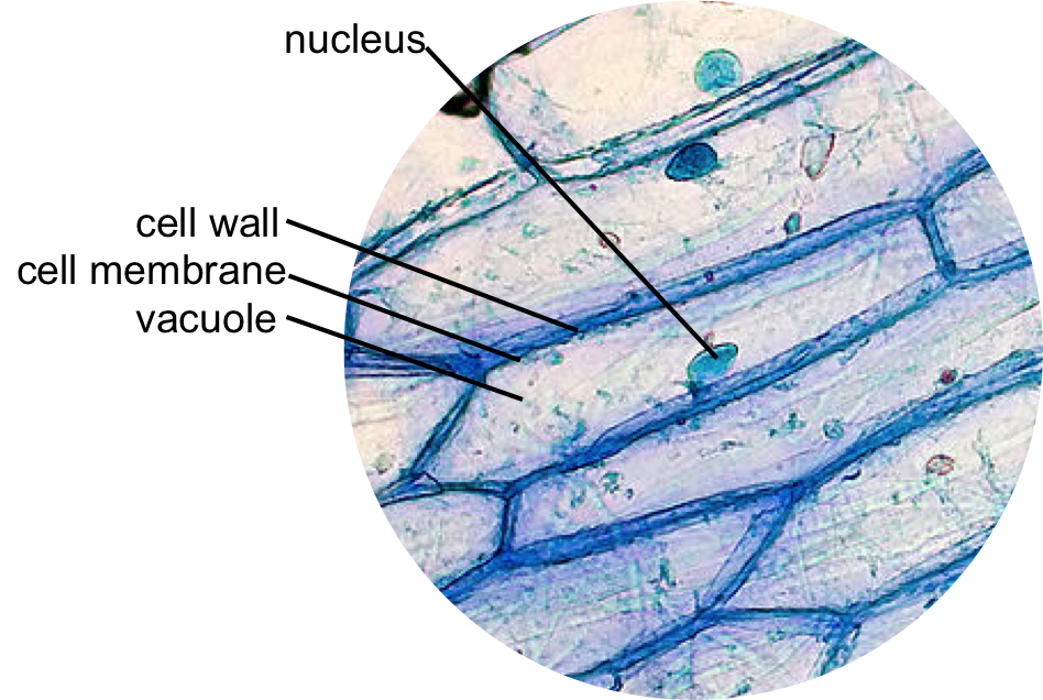
animal cell under light microscope
To use a light microscope to examine animal or plant cells. It directs all of the cells activities.
Under the microscope animal cells appear different based on the type of the cell.

. See also what does ostracise mean Do animal cells have a chloroplast. 2There are light-illuminated compound microscopes. It was not until good light microscopes became available in the early part of the nineteenth century that all plant and animal tissues were discovered to be aggregates of individual cells.
Hence the correct answer is option C Note. Most animal cells are between 001 mm 005 mm most plant cells are between. Made using Microsoft SP4 MovieMaker.
To prepare animal cells for viewing under a microscope Use a cotton swab to get cheek cells and smear this onto a slide. A typical animal cell is 1020 μm in diameter which is about one-fifth the size of the smallest particle visible to the naked eye. All of the specimens where viewed under x100 magnification this is achieved by the lenses on the eye piece producing x10 and then the objective lenses producing an additional x10 with in turn give x100 magnification.
The nucleus cytoplasm cell membrane chloroplasts and cell wall are organelles which can be seen under a light microscope. For example mitochondria cannot be seen with a standard lab light microscope. Animal Cell as shown above Plant cell as shown above The Cell as Seen under the Electron Microscope.
Light microscopes Greg Foot explains the main differences between light and electron microscopes Cells range in size. 14 can you distinguish features of the cells in Fig. Aims of the experiment.
Almost all animals and plants are made up of cells. Rinse off the stain and allow the slide to dry. Some of the cell organelles that can be observed under the light microscope include the cell wall cell membrane cytoplasm nucleus vacuole and chloroplasts.
To see the cell organelles you will need to get a higher magnification usually with a 40x-100x objective lens. These cell organelles perform specific functions within the cell. Once slides have been prepared they can be examined under a microscope.
Using a light microscope. A cell is the structural and functional unit of life. Microscope observation of animal and plant.
16 p4 which is photomicrograph of actual animal cells. Draw and label the structure of a generalized animal cell ie. Every organism composed of one or more cells.
Investigating cells with a light microscope. Aims of the experiment. All cells are categorized in to two groups- Prokaryotic and Eukaryotic.
In addition the electron microscope is required to resolve the structure of mitochondria bacteria viruses and large protein complexes. You can see a variety of cells pretty well with the light microscope. Below the basic structure is shown in the same animal cell on the left viewed with the light microscope.
For this sort of microscope the picture. This shows a generalized animal cell under a light microscope. Observing a wide range of biological processes and animal cell under light microscope is easier due to advances in microscopic techniques.
Human cheek cells are made of simple squamous epithelial cells which are flat cells with a round visible nucleus that cover the inside lining of the cheekC. Animal cells have a basic structure. The nucleus is the control center.
Using computational methods multiple images and views from different directions are. Once slides have been prepared they can be examined under a microscope. Using the labels of Fig.
Has 5 visible organelles looking through a compound light microscope. To use a light microscope to examine animal or. Under a light microscope mitochondria are still visible but thorough research.
Plant and Animal Cell Under a Compound Light Microscope. In this chapter we are making user to control a light microscope remotely using a eukaryotic cell. Put a couple of drops of methylene blue stain on the smear and leave for a few mintues.

List Of Cell Organelles Their Functions Plant And Animal Cells Animal Cell Cell Theory

Cells Under Electron Microscope Google Search Animal Cell Structure Animal Cells Worksheet Cell Diagram

Animal Cell Structure And Organelles With Their Functions

An Electron Micrograph Of A Mouse Liver Cell Dna Learning Center Electrons Cell Learning Centers

Onion Epidermis Light Microscope Purple Colored Large Epidermal Cells Onion

Onion Epidermis With Large Cells Under Light Microscope Stock Photo Image Of Layer Nucleus 88787098

Onion Epidermis Under Light Microscope Purple Colored Large Epidermal Cells Of An Onion Alli Things Under A Microscope Cells Project Microscopic Photography

Animal Cell Color Code The Organelles Animal Cell Color Coding Organelles

Living And Dyeing Under The Big Sky Cell Structure Images Cell Structure Microscopic Images Microscopic Photography

Onion Epidermis Under Light Microscope Purple Colored Large

Plant Cell Under The Microscope 1 Microscopic Photography Plant Cell Microscopic Images

Cell Theory Plant Cell Diagram Cell Diagram

1 5 Microscopy Biology Libretexts Microscopy Eukaryotic Cell Animal Cell

Plant Animal Cells Staining Lab Answers

Lab The Cell The Biology Primer

Cell Organelles And Their Function

Typical Animal And Plant Cells Sec Individual Microscope Slide


